Brain and ENT Clinic – Dr Lalit Mahajan In Nagpur & Dr Rachna Gangwani Mahajan In Nagpur
Electroencephalography EEG
Electroencephalography (EEG) is a non-invasive neurophysiological test that measures and records the electrical activity of the brain. It is commonly used to assess brain function and diagnose various neurological conditions. The EEG recording represents the collective electrical activity generated by the firing of neurons (brain cells) in the cerebral cortex.
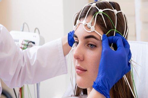
Small metal electrodes are attached to the scalp using a conductive gel or paste. These electrodes are strategically placed at specific locations to capture electrical signals from different regions of the brain.The electrical activity of the brain is recorded in the form of brain waves. These brain waves are categorized into different frequency bands, each associated with different states of consciousness, attention, and relaxation.
Reasons why EEG is Needed
Diagnosis of Epilepsy:
- EEG is a crucial tool for diagnosing and characterizing epilepsy. It helps identify abnormal electrical activity in the brain associated with seizures. The specific patterns and locations of abnormal activity can provide insights into the type and origin of seizures.
Evaluation of Seizure Disorders:
- Beyond epilepsy diagnosis, EEG is used to evaluate patients with seizure disorders to monitor the frequency and characteristics of seizures. It aids in adjusting treatment plans and assessing the effectiveness of anti-seizure medications.
Sleep Disorders Assessment:
- EEG is widely used in sleep medicine to assess sleep stages and identify abnormalities during sleep. It helps diagnose conditions such as insomnia, sleep apnea, and parasomnias.
Monitoring Brain Function during Surgery:
- In certain neurosurgical procedures, especially those involving the brain’s functional areas, EEG is used to monitor brain activity in real-time. This helps surgeons avoid critical areas responsible for motor function, language, and sensory perception.
Research in Neuroscience:
- EEG is a valuable tool in neuroscience research, providing insights into brain function during various cognitive tasks, perception, memory, and emotion.
Steps involved in conducting an EEG Procedure
1. Preparation:
- The patient is typically instructed to wash their hair before the EEG to ensure a clean scalp, free of oils and hair products. It’s important to inform the healthcare provider about any medications the patient is taking, as certain medications may need to be temporarily stopped before the test.
2. Placement of Electrodes:
- The EEG technician, often a neurophysiologist or EEG technologist, will measure the patient’s head and use a small amount of conductive gel to attach electrodes to specific locations on the scalp. The number and placement of electrodes can vary, but a standard EEG setup typically involves placing electrodes in a specific pattern, such as the International 10-20 system.
3. Recording Resting State:
- The patient is usually asked to sit or lie down in a comfortable position. During the resting state, the EEG records the baseline electrical activity of the brain. The patient may be instructed to close their eyes or keep them open, depending on the specific goals of the test.
4. Hyperventilation:
- In some cases, the patient may be asked to hyperventilate by breathing rapidly and deeply for a short period. This can trigger certain abnormalities in the EEG, helping in the diagnosis of conditions like epilepsy.
5. Photic Stimulation:
- Photic stimulation involves exposing the patient to flickering lights at different frequencies. This can be done using a strobe light or a special device. Photic stimulation may be used to elicit specific responses in the EEG, particularly in the diagnosis of certain seizure disorders.
6. Sleep Deprivation:
- In some cases, the patient may be asked to deprive themselves of sleep the night before the EEG. Sleep deprivation can increase the likelihood of capturing abnormal brain activity during drowsiness or sleep.
7. Video Monitoring:
- Many EEG recordings are done in conjunction with video monitoring. This allows healthcare providers to correlate the EEG findings with the patient’s behavior, movements, and any visible manifestations of seizures.
8. Recording Duration:
- The duration of an EEG recording can vary. Routine EEGs typically last 20 to 60 minutes, but in some cases, especially during long-term monitoring or video EEG studies, the recording may continue for several hours or even days.
9. Removal of Electrodes:
- Once the recording is complete, the electrodes are removed, and any remaining gel is cleaned from the scalp. Patients can resume their normal activities after the procedure.
10. Interpretation: – The recorded EEG data is analyzed by a neurologist or a clinical neurophysiologist. They interpret the findings, looking for patterns or abnormalities that may indicate various neurological conditions.